Identificación molecular de la microbiota intestinal cultivable de Epinephelus morio (Mero rojo) y su potencial probiótico: Microbiota intestinal del Mero rojo Ephinephelus morio.

Publicado 2025-07-01
Palabras clave
- Bacteria,
- microbiota intestinal cultivable,
- peces marinos,
- potencial probiótico de mero rojo,
- secsuenciación genética 16S rRNA
Cómo citar
Derechos de autor 2025 Maria Leticia Arena Ortiz, Montalvo-Fernandez Grecia, Ortíz-Alcántara Joanna, Durruty-Lagunes Claudia

Esta obra está bajo una licencia internacional Creative Commons Atribución-NoComercial-CompartirIgual 4.0.
Resumen
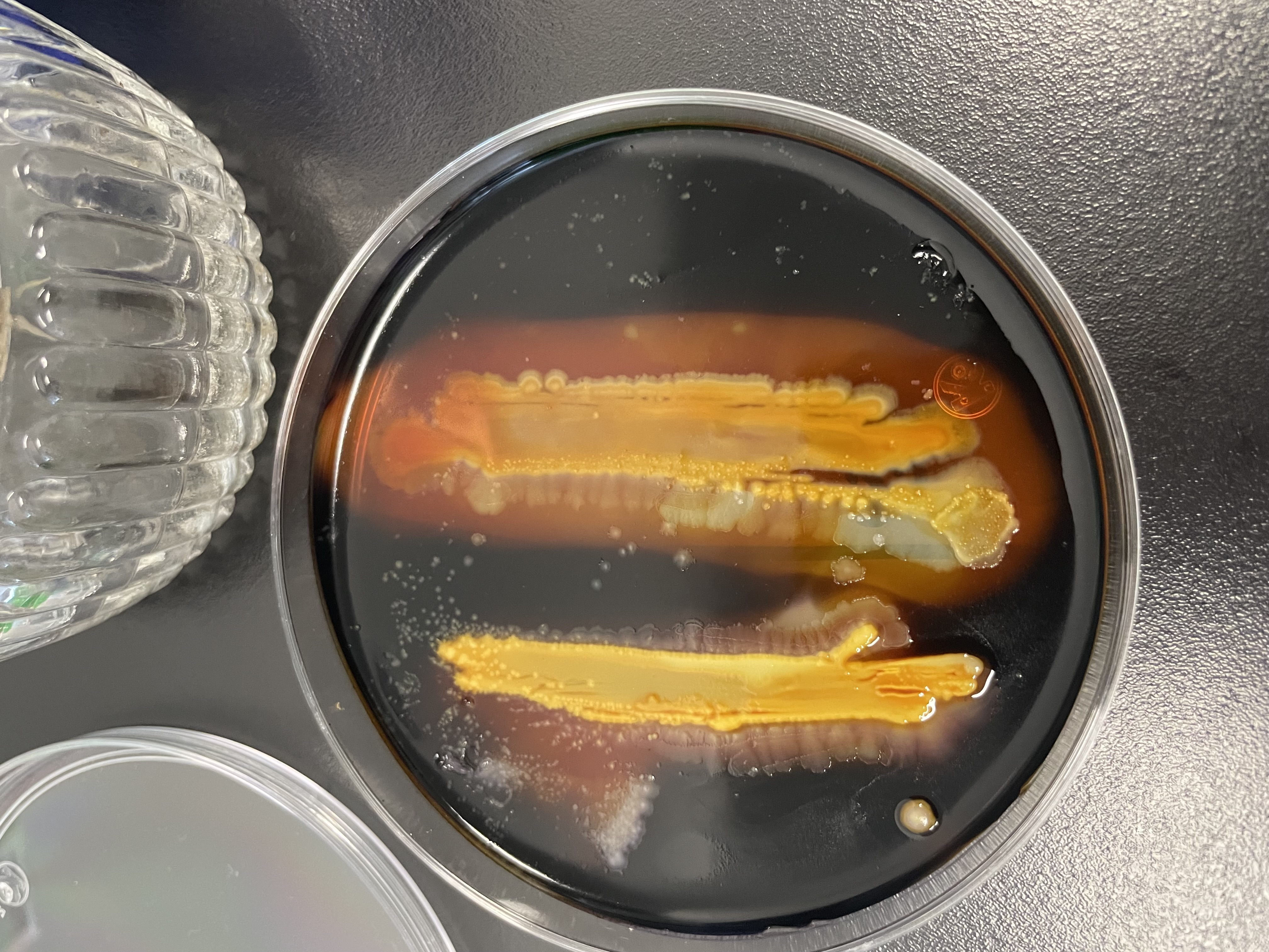
La microbiota intestinal de Epinephelus morio (Mero rojo) es importante para la digestión, metabolismo y salud. Sin embargo, se sabe poco sobre la composición de la microbiota en esta especie y en cautiverio podría verse afectada por la composición de la dieta y otros factores ambientales. En este estudio, la microbiota intestinal cultivable del mero rojo silvestre, mantenido en cautiverio, se caracterizó mediante la secuenciación del gen ARNr 16S y se estudió su potencial como probiótico. Se utilizaron 6 peces adultos entre 3-7 kg y se tomó el contenido intestinal mediante una sonda. Las muestras se cultivaron en Agar Soya Trpticaseína y Agar Marino. La obtención de aislados bacterianos se llevó a cabo usando los mismos medios de cultivo. Posteriormente se procedió a la extracción de DNA y amplificación del gen ARNr 16S. Los amplicones se purificaron y se enviaron a secuenciar por el método de Sanger. Para la identificación taxonómica se empleó la herramienta BLASTn de la plataforma NCBI. Se determinó la actividad amilolítica, quitinolítica y proteolítica de los aislados empleando medios de cultivo específicos. Se obtuvieron 22 aislados, 10 provenientes de medio marino y 12 de TSA. Los phyla que se identificaron fueron Proteobacteria, Firmicutes y Actinobacteria. La especie más representada fue Photobacterium damselae presente en el 31.8 % de las muestras. Dos aislados presentaron actividad amilolítica y 13 aislados tuvieron actividad proteolítica. Ninguno fue positivo para la actividad quitinolítica.
Descargas
Citas
- Abd El-Rhman, A.M., Khattab, Y.A.E. and Shalaby, A.M.E. (2009). Micrococcus luteus and Pseudomonas species as probiotics for promoting the growth performance and health of Nile tilapia, Oreochromis niloticus. Fish & Shellfish Immunology, 27(2), pp.175–180. https://doi.org/10.1016/j. fsi.2009.03.020.
- Abedin, M.Z., Rahman, S., Hasan, R., Shathi, J.H., Jarin, L. and Uz Zaman, S. (2020). Isolation, identification, and antimicrobial profiling of bacteria from aquaculture fishes in pond water of bangladesh. American Journal of Pure and Applied Biosciences, 2(3), pp.39–50. https://doi.org/10.34104/ajpab.020.039050.
- Aguilera, E., Yany, G. and Romero, J. (2013). Cultivable intestinal microbiota of yellow tail juveniles (Seriola lalandi) in an aquaculture system. Latin American Journal of Aquatic Research, 41(3), pp.395–403. https://doi.org/10.3856/vol41-issue3-fulltext-3.
- Alcántara-Jaúregui, F.M., Valladares-Carranza, B. and Ortega, C. (2022). Enfermedades bacterianas y sus agentes etiológicos identificados en peces de México - Una revisión. Revista MVZ Córdoba, 27(2), pp.1–12. https://doi.org/10.21897/rmvz.2387.
- Buruiană, C.T., Profir, A.G. and Vizireanu, C. (2014). Effects of probiotic Bacillus species
- in aquaculture. The Annals of the University Dunarea de Jos of GalatiFascicle VI – Food Technology, 38(2), pp.9–17. Available at: https://www.gup.ugal.ro/ugaljournals/index.php/food/article/view/1741/1470
- Butt, R.L. and Volkoff, H. (2019). Gut microbiota and energy homeostasis in fish. Frontiers in Endocrinology, 10. https://doi.org/10.3389/fendo.2019.00009.
- Cabra, T., Rodríguez, C.A. y Villota, C.P. (2014). Capacidad antagónica y quitinolítica de microorganismos aislados de residuos de higuerilla (Ricinus communis). Biotecnología en el Sector Agropecuario y Agroindustrial, 12(1), pp. 56–61. Available at: https://revistas.unicauca.edu.co/index.php/ biotecnologia/article/ view/328/523
- Cao, S., Geng, Y., Yu, Z., Deng, L., Gan, W., Wang, K., Ou, Y., Chen, D., Huang, X., Zuo, Z., He, M. and Lai, W. (2018). Acinetobacter lwoffii, an emerging pathogen for fish in Schizothorax genus in China. Transboundary and Emerging Diseas- es, 65(6), pp.1816–1822. https://doi.org/10.1111/ tbed.12957.
- Dehler, C.E., Secombes, C.J. and Martin, S.A.M. (2017). Seawater transfer alters the intestinal microbiota profiles of Atlantic salmon (Salmo salar L.). Scientific Reports, 7(13877), pp.1–11. https://doi.org/10.1038/s41598-017-13249-8.
- Dharmaraj, R. and Rajendren, V. (2014). Probiotic assessment of Bacillus infantis isolated from gastrointestinal tract of Labeo rohita. International Journal of Scientific and Research Publications, 4(7), pp.1–6. Available at: https://www.ijsrp.org/research-paper-0714/ijsrp-p3188.pdf [Accessed 30 Apr. 2025].
- do Vale Pereira, G., da Cunha, D.G., Pedreira Mourino, J.L., Rodiles, A., Jaramillo-Tor- res, A. and Merrifield, D.L. (2017). Characterization of microbiota in Arapaima gigas intestine and isolation of potential probiotic bacteria. Journal of Applied Microbiology, 123(5), pp.1298–1311. https://doi.org/10.1111/jam.13572.
- Drider, D., Bendali, F., Naghmouchi, K. and Chikindas, M.L. (2016). Bacteriocins: Not only antibacterial agents. Probiotics and Antimicrobial Proteins, 8(4), pp.177–182. https:// doi.org/10.1007/s12602-016-9223-0.
- Egerton, S., Culloty, S., Whooley, J., Stanton, C. and Ross, R.P. (2018). The gut microbiota of marine fish. Frontiers in Microbiology, 9. https://doi.org/10.3389/ fmicb.2018.00873Felsenstein, J. (1985). Confidence limits on phylogenies: An approach using the bootstrap. Evolution, 39(4), pp.783–791. https://doi.org/10.1111/j.1558-5646.1985.tb00420.x.
- Gai, C., Liu, J., Zheng, X., Xu, L. and Ye, H. (2022). Identification of Vibrio ponticus as a bacterial pathogen of coral trout Plectropomus leopardus. Frontiers in Cellular and Infection Microbiology, 12. https://doi.org/10.3389/ fcimb.2022.1089247.
- Ghanbari, M., Kneifel, W. and Domig, K.J. (2015). A new view of the fish gut microbiome: Advances from next-generation sequencing. Aquaculture, 448(1), pp.464–475. https://doi.org/10.1016/j.aquaculture.2015.06.033.
- Hau, H.H. and Gralnick, J.A. (2007). Ecology and biotechnology of the genus Shewanella. Annual Review of Microbiology, 61, pp.287–308. https://doi.org/10.1146/annurev. micro.61.080706.093257.
- Kesarcodi-Watson, A., Kaspar, H., Lategan, M.J. and Gibson, L. (2008). Probiotics in aquaculture: The need, principles and mechanisms of action and screening processes. Aquaculture, 274(1), pp.1–14. https://doi.org/%2010.1016/j.aquacul- ture.2007.11.019.
- Kim, Y.-R., Kim, E.-Y., Choi, S., Hossain, M.T., Oh, R., Heo, W.- S., Lee, J.M., Cho, Y.C. and Kong, I.-S. (2012). Effect of a Probiotic strain, Enterococcus faecium, on the immune responses of olive flounder (Paralichthys olivaceus). Journal of Microbiology and Biotechnology, 22(4), pp.526–529. https://doi.org/10.4014/ jmb.1108.08047.
- Kim, P.S., Shin, N.-R., Lee, J.-B., Kim, M.-S., Whon, T.W., Hyun, D.-W., Yun, J.-H., Jung, M.-J., Kim, J.Y. and Bae, J.-W. (2021). Host habitat is the major determinant of the gut microbiome of fish. Microbiome, 9(1). https://doi.org/10.1186/s40168- 021-01113-x.
- Kozińska, A., Paździor, E., Pękala, A. and Niemczuk, W. (2014). Acinetobacter johnsonii and Acinetobacter lwoffii - the emerging fish pathogens. Journal of Veterinary Research, 58(2), pp.193–199. https://doi.org/10.2478/bvip-2014- 0029.
- Kuebutornye, F.K.A., Abarike, E.D. and Lu, Y. (2019). A review on the application of Bacillus as probiotics in aquaculture. Fish & Shellfish Immunology, 87, pp.820–828. https://doi.org/10.1016/j.fsi.2019.02.010.
- Liu, H., Guo, X., Gooneratne, R., Lai, R., Zeng, C., Zhan, F. and Wang, W. (2016). The gut microbiome and degradation enzyme activity of wild freshwater fishes influenced by their trophic levels. Nature. Scientific Reports, 6(24340), pp.1–12. https://doi.org/10.1038/srep24340.
- Llewellyn, M.S., Boutin, S., Hoseinifar, S.H. and Derome, N. (2014). Teleost microbiomes: the state of the art in their characterization, manipulation and importance in aquaculture and fisheries. Frontiers in Microbiology, 5, pp.1–17. https://doi. org/10.3389/fmicb.2014.00207.
- López Nadal, A., Ikeda-Ohtsubo, W., Sipkema, D., Peggs, D., McGurk, C., Forlenza, M., Wiegertjes, G.F. and Brugman, S. (2020). Feed, microbiota, and gut immu- nity: Using the zebrafish odel to understand fish fealth. Frontiers in Immunology, 11. https://doi.org/10.3389/fimmu.2020.00114.
- Magariños, B., Couso, N., Noya, M., Merino, P., Toranzo, A.E. and Lamas, J. (2001). Effect of temperature on the development of pasteurellosis in carrier gilthead seabream (Sparus aurata). Aquaculture, 195(1-2), pp.17–21. https://doi.org/10.1016/S0044-8486(00)00547-0.
- Merrifield, D.L. and Rodiles, A. (2015). The fish microbiome and its interactions with mucosal tissues. In: B.H. Beck and E. Peatman, eds., Mucosal Health in Aquaculture. Elsevier, pp.273–295. https://doi.org/10.1016/B978-0-12-417186- 2.00010-8.
- Musharrafieh, R., Tacchi, L., Trujeque, J., LaPatra, S. and Salinas, I. (2014). Staphylococ cus warneri, a resident skin commensal of rainbow trout (Oncorhynchus mykiss) with pathobiont characteristics. Veterinary Microbiology, 169(1-2), pp.80-88. https://doi.org/10.1016/j.vetmic.2013.12.012.
- Navarrete, P., Espejo, R.T. and Romero, J. (2008). Molecular analysis of microbiota along the digestive tract of juvenile Atlantic salmon (Salmo salar L.). Microbial Ecology, 57(3), pp.550–561. https://doi.org/10.1007/s00248-008-9448-x.
- Navarrete, P., Magne, F., Mardones, P., Riveros, M., Opazo, R., Suau, A., Pochart, P. and Romero, J. (2010). Molecular analysis of intestinal microbiota of rainbow trout (Oncorhynchus mykiss). FEMS Microbiology Ecology, 71(1), pp.148–156. https://doi.org/10.1111/j.1574-6941.2009.00769.x.
- Navarrete, P., Magne, F., Araneda, C., Fuentes, P., Barros, L., Opazo, R., Espejo, R. and Romero, J. (2012). PCR-TTGE Analysis of 16S rRNA from rainbow trout (Oncorhynchus mykiss) gut microbiota reveals host-specific communities of active bacteria. PLoS One, 7(2), p.e31335. https:// doi.org/10.1371/journal. pone.0031335.
- Nayak, S.K. (2010). Role of gastrointestinal microbiota in fish. Aquaculture Research, 41(11), pp.1553–1573. https://doi.org/10.1111/j.1365-2109.2010.02546.x.
- Ou, W., Yu, G., Zhang, Y. and Mai, K. (2021). Recent progress in the understanding of the gut microbiota of marine fishes. Marine Life Science & Technology, 3(4), pp.434–448. https://doi.org/10.1007/s42995-021-00094-y.
- Perry, W.B., Lindsay, E., Payne, C.J., Brodie, C. and Kazlauskaite, R. (2020). The role of the gut microbiome in sustainable teleost aquaculture. Proceedings of the Royal Society B: Biological Sciences, 287(1926), p.20200184. https://doi.org/10.1098/rspb.2020.0184.
- Reda, R.M., Selim, K.M., El-Sayed, H.M. and El-Hady, M.A. (2017). In vitro selection and identification of potential probiotics isolated from the gastrointestinal tract of Nile tilapia, Oreochromis niloticus. Probiotics and Antimicrobial Proteins, 10(4), pp.692–703. https://doi.org/10.1007/ s12602-017-9314-6.
- Rivas, A.J., Lemos, M.L. and Osorio, C.R. (2013). Photobacterium damselae subsp. damselae, a bacterium pathogenic for marine animals and humans. Frontiers in Microbiology, 4, pp.283–289. https://doi.org/10.3389/fmicb.2013.00283.
- Saitou, N. and Nei, M. (1987). The neighbor-joining method: a new method for reconstructing phylogenetic trees. Molecular Biology and Evolution, 4(4), pp.406–425. https://doi.org/10.1093/oxfordjournals.molbev.a040454.
- Selim, K.M. and Reda, R.M. (2015). Improvement of immunity and disease resistance in the Nile tilapia, Oreochromis niloticus, by dietary supplementation with Bacillus amyloliquefaciens. Fish & Shellfish Immunology, 44(2), pp.496–503. https:// doi.org/10.1016/j.fsi.2015.03.004.
- Serra, C.R., Almeida, E.M., Guerreiro, I., Santos, R., Merrifield, D.L., Tavares, F., Oliva- Teles, A. and Enes, P. (2019a). Selection of carbohydrate-active probiotics from the gut of carnivorous fish fed plant-based diets. Scientific Reports, 9(1), pp.1–15. https://doi.org/10.1038/s41598-019-42716-7.
- Serra, C.R., Almeida, E.M., Guerreiro, I., Santos, R., Merrifield, D.L., Tavares, F., Oliva-Teles, A. and Enes, P. (2019b). Selection of carbohydrate-active probiotics from the gut of carnivorous fish fed plant-based diets. Scientific Reports, 9(1), pp.1–15. https://doi.org/10.1038/s41598-019-42716-7.
- Smriga, S., Sandin, S.A. and Azam, F. (2010). Abundance, diversity, and activity of microbial assemblages associated with coral reef fish guts and feces. FEMS Microbiology Ecology, 73(1), pp.31–42. https://doi.org/10.1111/j.1574-6941.2010.00879.x.
- Sun, Y., Yang, H., Ling, Z., Chang, J. and Ye, J. (2009). Gut microbiota of fast and slow growing grouper Epinephelus coioides. African Journal of Microbiology Research, 3, pp.713–720. Available at: https://academicjournals.org/article/ article1380369856_Yunzhang.pdf [Accessed 7 May 2025].
- Sun, Y.Z., Yang, H.L., Ma, R.L., Zhang, C.X. and Lin, W.Y. (2011). Effect of dietary administration of Psychrobacter sp. on the growth, feed utilization, digestive enzymes and immune responses of grouper Epinephelus coioides. Aquaculture Nutrition, 17(3), pp.733–740. https://doi.org/10.1111/j.1365-2095.2010.00837.x.
- Suresh, K., Pillai, D., Soni, M., Rathlavath, S. and Narshivudu, D. (2024). Micrococcus luteus, an emerging opportunistic pathogen in farmed Nile tilapia, Oreochromis niloticus in Andhra Pradesh, India. Aquaculture International, 33(51). https://doi.org/10.1007/s10499-024-01761-4.
- Tamura, K. and Nei, M. (1993). Estimation of the number of nucleotide substitutions in the control region of mitochondrial DNA in humans and chimpanzees. Molecular Biology and Evolution, 10(3). https://doi:%2010.1093/oxfordjournals.molbev. a040023.
- Tamura, K., Stecher, G. and Kumar, S. (2021). MEGA11: Molecular Evolutionary Genetics Analysis Version 11. Molecular Biology And Evolution, 38(7), pp.3022–3027. https://doi.org/10.1093/molbev/msab120.
- Tapia-Paniagua, S.T., Vidal, S., Lobo, C., Prieto-Álamo, M.J., Jurado, J., Cordero, H., Cerezuela, R., García de la Banda, I., Esteban, M.A., Balebona, M.C. and Moriñigo, M.A. (2014). The treatment with the probiotic Shewanella putrefaciens Pdp11 of specimens of Solea senegalensis exposed to high stocking densities to enhance their resistance to disease. Fish & Shellfish Immunology, 41(2), pp.209–221. https://doi.org/10.1016/j.fsi.2014.08.019.
- Tortora, G., Funke, B.R. y Case, C.L. (2007). Introducción a la microbiología. Buenos Aires: Médica Panamericana, p.959.
- Wang, A.R., Ran, C., Ringø, E. and Zhou, Z.G. (2018). Progress in fish gastrointestinal microbiota research. Reviews in Aquaculture, 10(3), pp.626–640. https://doi.org/10.1111/raq.12191.
- Xiao Joe, J.T., Tseng, Y.-C., Wu, J.-L. and Lu, M.-W. (2021). The alteration of intestinal microbiota profile and immune response in Epinephelus coioides during pathogen infection. Life, 11(2), p.99. https://doi.org/10.3390/life11020099.
- Yarza, P., Yilmaz, P., Pruesse, E., Glöckner, F.O., Ludwig, W., Schleifer, K.-H., Whitman, W.B., Euzéby, J., Amann, R. and Rosselló-Móra, R. (2014). Uniting the classification of cultured and uncultured bacteria and archaea using 16S rRNA gene sequences. Nature Reviews Microbiology, 12(9), pp.635–645. https://doi.org/10.1038/nrmicro3330.
- Zatán Valdiviezo, A.E., Castillo Chunga, D., Castañeda Vargas, A.E., Feria Zevallos, M.A., Toledo Valdiviezo, O.E., Aguilar Zavaleta, J.L., Cueva Távara, M.D. and Motte, E. (2020). Characterization of the intestinal microbiota in snook (Centropomus sp.) and isolation of potential probiotic bacteria. Revista de Investigaciones Veterinarias del Perú, 31(3), p.e16036. https://doi.org/10.15381/rivep. v31i3.16036.
- Zhang, B., Yang, H., Cai, G., Nie, Q. and Sun, Y. (2024). The interactions between the host immunity and intestinal microorganisms in fish. Applied Microbiology and Biotechnology, 108(30), pp.1–14. https://doi.org/10.1007/s00253-023-12934-1.